Breaking the diffraction barrier
A recent paper from Stefan W. Hell group (Nature 440, 935-939, 2006) uses stimulation emission depleted microscopy - STED microscopy, to address the faith of synaptotagmin I, a vesicle protein, after stimulation of neurotransmitter release. This technology allows for resolutions well bellow the theoretical diffraction resolution limits of light microscopy (down to ~60nm).
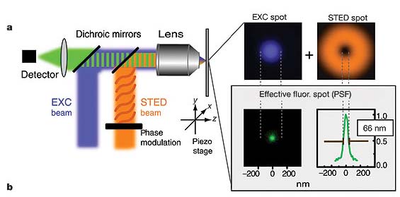
"In a typical STED microscope the excitation beam is overlapped with a doughnut-shaped beam that is capable of de-exciting fluorophores by stimulated emission. Co-alignment of the beams ensures that fluorescence is allowed only in the central area of the excitation spot where the doughnut beam is close to zero" - see figure right
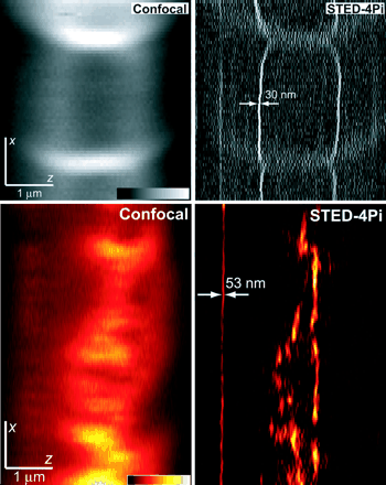
This is not the only current available technology where the diffraction barrier of visible light has been beaten. Others approaches such as "4Pi", "I3M" and "I5M" are also used. One key aspect of these technologies is the resolution which is obtained in the Z-axis, much better than any available confocal microscope - see figure left. This will revolutionize fields such as epithelial cell polarity.
Currently only Leica (Leica TCS 4PI) sells microscopes with this technology, although soon other companies will release equivalent products.
Further reading:
Blinded by the Light, Bio-IT world
Hell, S. W. Toward fluorescence nanoscopy. Nature Biotechnol. 21, 1347Â1355 (2003)
posted by lost @ 5:30 PM
0 comments
![]()

0 Comments:
Post a Comment
<< Home