Something in the tubes, Part 2
Biology is first and foremost driven by observations. Much of the current field of cell biology involves working out the details of phenomena that were observed a hundred years ago or more using the naked eye, shaped glass, and rudimentary microscopes. Today's biologists are armed with ever more powerful tools to peer deeper into the molecular level of the cell and see things we had never seen before. Even subtle changes in the methods of preparing samples enable us to see things we hadn't previously seen. The paper I highlight today is one such example.
Like yesterday, I am presenting an electron microscopy study of axonemal microtubules. Yesterday's paper was the flagellum of sea urchin sperm; today's is the flagellum of the African trypansome Trypanosoma brucei (responsible for African sleeping sickness). Although the two organisms are separated by millions of years of evolution, the inherent ultrastructure of their flagella are the same (another example of nature's elegant simplicity) - but I digress. Previously, tannic acid was used in the fixation of samples for transmission electron microscopy (a relatively simpler technique than yesterday's cryo-EM study) to enhance the contrast of the resulting image (as seen in panel B of the figure below compared to panel A). However, Vaughan and colleagues noted that in the absence of tannic acid a structure is retained in the lumen of the B-tubule of the microtubule doublet that is not apparent in the presence of tannic acid. Here's the image:
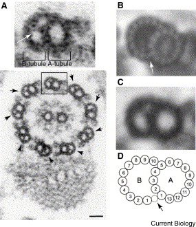
They call this structure the "ponticulus" (little bridge) and note that it is present in all nine of the outer doublet microtubules of the flagellum (see lower portion of panel A, above). They go on to point out that newly formed flagella do not contain this structure, but that it is instead modified later - after the flagellum has formed. They also note (although no data is shown) that this structure is also present in the flagellum of other trypanosomes, namely Leishmania and Chrithidia.
What is this ponticulus? Since it is formed after the flagellum is made, how does it get access to the outer tubule of the doublet? Is it necessary for the structural integrity of the trypanosome flagellum? Also, why was it not seen in the study by Sui and Downing of sea urchin spem flagella (see below)? Is it unique to trypanosomes? Like early observations of biological phenomena, these observations will drive future work in cell biology.
Reference:
Vaughan, S., Shaw, M., and Gull, K. (2006). A post-assembly structural modification to the lumen of flagellar microtubule doublets. Curr. Biol. 16, R449-50.
posted by Anonymous @ 11:37 AM
1 comments
![]()

1 Comments:
Man, you're really into this MT breathing thing.
Post a Comment
<< Home