5.30.2006

Three Brief Papers on Nuclear Pore Complexes

The nuclear membrane separates the nuclear space from the cytoplasm. This barrier is comprised of two membranes (Inner and Outer Nuclear Membrane) that are continuous with the endoplasmic reticulum. To cross the double membrane, molecules traverse the nuclear pore complex (NPC), a giant macromolecular complex that has an eight-fold symmetry and weighs over 100MDa. To date, only two components of the NPC have been identified: gp210 and Pom120. Interestingly many cells only express one of these proteins. Well it seems like Dirk Gorlich’s group have been able to knock out BOTH genes from Hela cells … and the cells are fine! They have NPCs, they can reform the nuclear membrane after mitosis … something strange is going on (On top of that another group previously claimed that gp210 was essential … in Hela cells!)

Nuclear pore complex assembly and maintenance in POM121- and gp210-deficient cells
Fabrizia Stavru, Gitte Nautrup-Pedersen, Volker C. Cordes, and Dirk Görlich
JCB (2006) 173:477-83


In a second paper the Gorlich group solve the problem … there is a third integral membrane protein that is part of the NPC, called Ncd1. Turns out that yeast don’t have gp210 or Pom120 orthologues. Instead they have Ncd1 and two other non-conserved integral membrane proteins, Pom152 and Pom34. Of the three, Ncd1 is the only protein that is essential and conserved in other organisms. Interestingly Ncd1 is also part of the yeast spindle pole body, a structure related to our centrosomes … (Does this sound interesting Gomez?). The Gorlich lab show that there are Ncd1 orthologues that localize to NPCs in fly, frogs, worms, humans

The Gorlich lab knocked down Ncd1 in Hela and the NPCs were defective (lower staining of NPC components). Worm knockouts were mostly inviable.

NDC1: a crucial membrane-integral nucleoporin of metazoan nuclear pore complexes Fabrizia Stavru, Bastian B. Hülsmann, Anne Spang, Enno Hartmann, Volker C. Cordes, and Dirk Görlich JCB (2006) 173:509-19

Last is a paper from Martin Hetzer’s lab. Here they tackle the tricky question of how do you form NPCs. There are two times when you have to form these structures; after mitosis when the nuclear envelope reforms, and during interphase as the nucleus expands. Using complicated in vitro manipulations of nuclei formed from frog egg extracts, the Hertzer lab shows that active Ran is required both in the nuclear and cytoplasmic compartments to activate NPC formation in an expanding nucleus (an analogous situation to NPC formation in interphase). They offer evidence that ran acts to dissociate the Nup107complex, which is a major constituent of the NPC) from Importin-beta in both the nucleoplasm and cytoplasm. Interestingly they show that excess free Nup107 complex can stimulate NPC formation IN NUCLEI THAT DO NOT HAVE NPCs. That means that exogenously added Nup107 complex can stimulate the fusion of the Outer Nuclear Membrane and Inner Nuclear Membrane, to form a hole where the NPC sits. They then observe that gp210 form patches on the nuclear envelope and may act as landing pads for Nup107 complex in intact nuclei.

What this last paper shows is that within the lumen of the nuclear envelope threre is some machinery that acts to fuse the two membranes to form pores. What is the machine? Well it looks like of the integral membrane proteins, gp210 and Pom120 are dispensable. Ncd1? There are survivors of the Ncd1 knockouts in worms. These survivors also have NPCs. Thus it is likely that a whole fusion complex exists but is waiting for someone to find it …

Nuclear Pores Form de Novo from Both Sides of the Nuclear Envelope
Maximiliano A. D’Angelo, Daniel J. Anderson, Erin Richard, Martin W. Hetzer
Science (2006) 312:440-443


Cross posted at the Daily Transcript.
5.23.2006

ER compartmentalization

The secretory membrane system in the Drosophila syncytial blastoderm embryo exists as functionally compartmentalized units around individual nuclei
David Frescas, Manos Mavrakis, Holger Lorenz, Robert DeLotto, and Jennifer Lippincott-Schwartz
J. Cell Biol. 2006 173: 219-230.

A very simple question was addressed in this work. Is the ER and Golgi compartmentalized in the Drosophila syncytial blastoderm embryo? The answer is NO and YES. NO, if nuclei are still in the interior of the embryo, and YES, if the nuclei are already in the periphery of the embryo. The authors also found that the compartmentalization is disrupted when microtubules are depolymerized after the nuclei arrived to the periphery. Also this compartmentalization occurs prior to cellularization, suggesting a role for the microtubule cytoskeleton on the formation of distinct ER and Golgi compartments within the same space.
Now the question is what are the mechanisms that give rise to this compartmentalization.

note: during proof reading of this post, a comment on the same paper was published in this blog by the mad scientist. Any similarities between posts is a pure coincidence and should not be use against the authors of this blog.

Three Brief Papers on the ER

Here are some cool ER papers I've seen recently:

Direct membrane protein-DNA interactions required early in nuclear envelope assembly
Sebastian Ulbert, Melpomeni Platani, Stephanie Boue, and Iain W. Mattaj
JCB (2006) 173:469-476


When the nuclear envelope reforms after mitosis, ER vesicles must bind to the condensed chromatin, but how does this occur? Well about half of the nuclear envelope (NE) proteins have basic luminal domains that mediate electrostatic interactions with the DNA itself. (In comparison about 4% of general ER and Golgi proteins have basic luminal domains.) To prevent ER/DNA association during mitosis, these basic NE proteins are phosphorylated.

ER-bound PTP1B is targeted to newly forming cell-matrix adhesions
Mariana V. Hernández, Maria G. Davies Sala, Janne Balsamo, Jack Lilien and Carlos O. Arregui
JCS (2006) 119:1233-1243


How does the ER remain extended in cells? This becomes a problem once you realize that actin, which is constantly polymerized at the cell's edge, is constantly being pushed towards the nucleus, and taking everything else along with it. Now it seems like the ER can interact with focal adhesions via protein tyrosine phosphatase 1B. This interaction may help anchor the ER at peripheral focal adhesion sites. Whether this is the only ER protein that can interact with focal adhesions remains unclear.

The secretory membrane system in the Drosophila syncytial blastoderm embryo exists as functionally compartmentalized units around individual nuclei
David Frescas, Manos Mavrakis, Holger Lorenz, Robert DeLotto, and Jennifer Lippincott-Schwartz
JCB (2006) 173:219-230


In Drosophila melanogaster embryogenesis, the first 13 nuclear division cycles are not accompanied by any cellularization. What you get is a giant single syncytium with over 6000 nuclei. At a specific time point all the nuclei migrate to the surface of the syncytium and then around each nuclei a cell membrane is constructed. In this paper the authors examined how the ER and Golgi of the syncytium are formed. After nuclear migration, the ER which previously was one giant cortical network, compartmentalizes around each nucleus. This even occurs prior to cellularization and requires a functional microtubule array.

5.08.2006

Lamins are cool!!!

Lamin A-Dependent Nuclear Defects in Human Aging

Paola Scaffidi 1 and
Tom Misteli 1*
Published online April 27 2006; 10.1126/science.1127168 (Science Express Reports)

Lamins are intermediate filament proteins and are the main component of the nuclear lamina. Although these proteins are expressed in different tissues, mutations in the LMN gene that encodes Lamin A and Lamin C, are associated with distinct genetic disorders such as muscular dystrophies, lypodystrophies and progerias.

Now it was found that the same mutation on lamin A protein associated with progeria, a rare disease were children suffer from premature aging, also accumulates in elderly human cells. This mutation originates a truncated version of lamin A.

Two key findings are reported:
- Traces of this truncated version are also observed in cells from young humans, although the protein does not accumulate, as observed in elderly human cells. This suggests that younger cells have mechanisms to destroy this truncated version.
- Cell nuclei from elderly humans have the same morphology and DNA-damage levels as cell nuclei from progeria patients. Impressively, this morphology is reversed when the production of the truncated protein is prevented, by expression of a morpholino targeting the "truncated mRNA".

Maybe in the future cell aging can be prevented by inhibiting the formation of truncated lamin A.... It will be cool to generate a trangenic mice expressing the morpholino that inhibits the formation of spontaneous truncated lamin A.

5.07.2006

Breaking the diffraction barrier

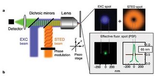
A recent paper from Stefan W. Hell group (Nature 440, 935-939, 2006) uses stimulation emission depleted microscopy - STED microscopy, to address the faith of synaptotagmin I, a vesicle protein, after stimulation of neurotransmitter release. This technology allows for resolutions well bellow the theoretical diffraction resolution limits of light microscopy (down to ~60nm).

"In a typical STED microscope the excitation beam is overlapped with a doughnut-shaped beam that is capable of de-exciting fluorophores by stimulated emission. Co-alignment of the beams ensures that fluorescence is allowed only in the central area of the excitation spot where the doughnut beam is close to zero" - see figure right


This is not the only current available technology where the diffraction barrier of visible light has been beaten. Others approaches such as "4Pi", "I3M" and "I5M" are also used. One key aspect of these technologies is the resolution which is obtained in the Z-axis, much better than any available confocal microscope - see figure left. This will revolutionize fields such as epithelial cell polarity.

Currently only Leica (Leica TCS 4PI) sells microscopes with this technology, although soon other companies will release equivalent products.


Further reading:
Blinded by the Light, Bio-IT world
Hell, S. W. Toward fluorescence nanoscopy. Nature Biotechnol. 21, 1347–1355 (2003)

Hi,

The idea of this blog came from long conversations with some other bloggers. Here is the deal: To describe what is currently being discussed in the 5 minute men journal club. What is the 5 min men? Me and some other people from Columbia Univ. get together every friday and present a paper in 5 min + 5 min discussion. We have been doing this for almost a year and it is great. Since one of the funding members is leaving to San Francisco, I thought on going virtual.

So I hope that this will work. Stay tuned.