8.27.2006

Inner Nuclear Membrane Proteins are Actively Imported

This is the newest from the Blobel lab.

Note to all "they've discovered everything" types: this finding shows how much we know about how cells operate.

Background: As I've described before the nucleus and the cytoplasm are two cellular compartments that are kept apart by the Nuclear Pore Complex (NPC). This mega-assembly of proteins is the gate (or the bouncer) of the nucleus - pass it and you can gain access to the nucleus from the cytoplasm (or vice versa). NPCs sit in the nuclear envelope, an extension of the ER that covers the chromosomes.

Nuclear proteins are synthesized in the cytoplasm. To cross the NPC and thus gain access to the nucleoplasm, these proteins are equiped with a nuclear localization sequence (NLS) that recruits special proteins called "Karyopherins" (alpha/beta importin in the diagram). Karyopherins are like VIPs, they can side step the bouncer and thus escort the proteins into the nucleus. Once inside, the importins release their guest, bind to the small G-protein Ran in it's GTP bound configuration, and are exported back out of the nucleus. Once out Ran hydrolyses its GTP into GDP + phosphate.

The whole import pathway is thus maintained by a Ran-GTP gradient across the membrane and requires energy (in the form of GTP).

But there are exceptions to this active import process. Smaller proteins (< 30 KDa) freely diffuse through the NPC, and it was thought that proteins found in the inner-nuclear-membrane could also diffuse across this barrier.

But now a new paper from the Blobel lab demonstrates that at least some inner-nuclear membrane proteins in yeast can only enter the nucleus by recruiting karyopherins. Since these karyopherins require a Ran-GTP gradient, this import process requires energy. So inner-nuclear-membrane proteins (in this diagram the red blob) may act just like most proteins - they can't just waltz into the nucleus without a VIP pass.

The NPC may not let just any old membrane protein into the inner-nuclear-envelope, and thus the composition of the inner-nuclear-membrane is more tightly regulated than we once believed. In other words this component of the ER (the inner-nuclear-membrane) may be more differentiated than we one thought.

Ref:

Megan C. King, C. Patrick Lusk and Günter Blobel
Karyopherin-mediated import of integral inner nuclear membrane proteins
Nature (06) AOP


Crossposted at The Daily Transcript.

8.22.2006

Something in the tubes, and Dynein!

A new paper from Dick McIntosh's group appeared in Science this week that is similar to the paper in Nature I highlighted last week. In fact, the title is almost identical (The Molecular Architecture of Axonemes Revealed by Cryoelectron Tomography vs. Molecular Architecture of Axonemal Doublets Revealed by Cryo-electron Tomography). However, in my opinion, the paper by the McIntosh group covers a great deal more ground than the paper by Downing's group. But, read them both and decide for yourself.

Flagella are composed of 9 microtubule doublets surrounding the central pair of single microtubules. Movement of flagella takes place as a result of pulling forces exerted by the microtubule-dependent motor, dynein. Nicastro et al. performed cryoelectron tomography of flagella from Chalmydomonas and sea urchin sperm and did 3D reconstruction of the entire network. The resolution level of their images of dynein provide insight not only into the localization of dynein relative to the microtubules, but also possible mechanisms of dynein activity as a result of the orientation of different parts of the dynein heavy chain. Summarizing models are shown below.

Of interest to me (you might have guessed) is that they also saw significant densities on the inner lumen of the microtubule doublets, which they call MIPs (Microtubule Inner Proteins). Three significant MIPs are observed in this study which all exhibit 8 or 16 nm periodicity along the MT lattice. They suggest that these unknown proteins may provide stability to the long-lived microtubules in the flagellum - just as taxol, which binds to the inner surface ot microtubules, also stabilizes microtubules. However, they make no strong statements as to the identity of these molecules.

I find it interesting to compare the results from the two EM studies (if you compare them side by side, flip one of them 180 degrees because they present their data in opposite orientations). Although they both picked up similar densities in the A-tubule, the McIntosh group does not see the density suggested to be Tektins by the Downing group. Conversely, the prominent density in the B-tubule seen by the McIntosh group is not reported in the work from the Downing group. Instead, they see a density on the other side of the B-tubule. Oddly, neither group see the "ponticulus" identified by Vaughan et al. Since they both used sea urchin sperm for at least part of their work, why wouldn't they see exactly the same thing? Someone more familiar with cryo-EM methods may be able to show where the two preparations are different. If that is true, does it mean that they both have incomplete models and there is more inside the MT lumen than either suggest individually?


LINK:
Nicastro, D., Schwartz, C., Pierson, J., Gaudette, R., Porter, M.E., and McIntosh, R. 2006. The Molecular Architecture of Axonemes Revealed by Cryoelectron Tomography. Science 313: 944-948.

8.21.2006

Rho and polarity establishment


Noah Jenkins, Jennifer R. Saam, and Susan E. Mango
Published online 27 July 2006, Science
[DOI: 10.1126/science.1130291]

Establishment of Cell Polarity is key in several cellular contexts. Small GTPases, in particular Cdc42, are known regulators of cell polarity. It was known from previous works that the sperm determines the posterior pole of the C. elegans embryo. Righ after fertilization, during pronuclear migration, a wave of cortical actin (cortical actin flow) and myosin II moves away from the site of sperm entry, which becomes the posterior pole, and is involved in polarization of PAR-3 and -6 to the anterior pole, and PAR-2 to the posterior.

The identity of the sperm protein responsible for setting the cue remained unanswered until now. Schonegg et al. identified a RHO-GAP (CYK-4) that is required for the establishment of polarity. CYK-4 is present in the sperm centrosome and no actin or myosin polarization is observed in the absence of paternal CYK-4. CYK-4 is a GAP for RHO, i.e. it inhibits RHO activation. The authors propose that CYK-4 breaks down the symmetry in the actin cytoskeleton, resulting in cortical actin flow away from sperm entry site.

So how does Rho regulates actin cytoskeleton? The obvious candidate is Rho Kinase (ROCK), which activates myosin II and originates actin bundling. So a model can be proposed where local inhibition of Rho by CYK-4, leads to inhibition of ROCK and myosin II at the future posterior pole. The C. elegans homologue of ROCK is Let-502 and it is not know to be involved in the establishment of C. elegans embryo polarity, so probably we will soon find out...

8.15.2006

Something in the tubes, Part 2

Biology is first and foremost driven by observations. Much of the current field of cell biology involves working out the details of phenomena that were observed a hundred years ago or more using the naked eye, shaped glass, and rudimentary microscopes. Today's biologists are armed with ever more powerful tools to peer deeper into the molecular level of the cell and see things we had never seen before. Even subtle changes in the methods of preparing samples enable us to see things we hadn't previously seen. The paper I highlight today is one such example.

Like yesterday, I am presenting an electron microscopy study of axonemal microtubules. Yesterday's paper was the flagellum of sea urchin sperm; today's is the flagellum of the African trypansome Trypanosoma brucei (responsible for African sleeping sickness). Although the two organisms are separated by millions of years of evolution, the inherent ultrastructure of their flagella are the same (another example of nature's elegant simplicity) - but I digress. Previously, tannic acid was used in the fixation of samples for transmission electron microscopy (a relatively simpler technique than yesterday's cryo-EM study) to enhance the contrast of the resulting image (as seen in panel B of the figure below compared to panel A). However, Vaughan and colleagues noted that in the absence of tannic acid a structure is retained in the lumen of the B-tubule of the microtubule doublet that is not apparent in the presence of tannic acid. Here's the image:

They call this structure the "ponticulus" (little bridge) and note that it is present in all nine of the outer doublet microtubules of the flagellum (see lower portion of panel A, above). They go on to point out that newly formed flagella do not contain this structure, but that it is instead modified later - after the flagellum has formed. They also note (although no data is shown) that this structure is also present in the flagellum of other trypanosomes, namely Leishmania and Chrithidia.

What is this ponticulus? Since it is formed after the flagellum is made, how does it get access to the outer tubule of the doublet? Is it necessary for the structural integrity of the trypanosome flagellum? Also, why was it not seen in the study by Sui and Downing of sea urchin spem flagella (see below)? Is it unique to trypanosomes? Like early observations of biological phenomena, these observations will drive future work in cell biology.

Reference:
Vaughan, S., Shaw, M., and Gull, K. (2006). A post-assembly structural modification to the lumen of flagellar microtubule doublets. Curr. Biol. 16, R449-50.

8.14.2006

Something in the tubes

For my inaugural post in the blog, I wanted to share a paper that covers a topic that I am really excited about. It's something I became interested in as a graduate student hanging out in Gregg Gundersen's laboratory and hope to spend some time on now that I'm a post-doc. It gets at the fundamental difference between microtubules and the other cytoskeletal proteins (actin and intermediate filaments). Unlike the other two elements, microtubules (as their name implies) are indeed tubes. The protofilaments assemble in such a way as to create an inner core with a diameter of 16 nanometers (admitedly small but would support a globular protein somewhere in the range of 300 kiloDaltons). Although a great deal of work has gone into studying proteins that bind the outer surface of microtubules (motors, MAPs, plus-end tracking proteins, etc.), very few people are looking at the inside. A recent publication in the journal Nature may get a few more people thinking about it.

Haixin Sui and Kenneth Downing, working at the Lawrence Berkeley National Laboratory did a cryo-electron tomography (fancy high resolution imgaing) study of axonemal microtubules (the microtubules found in cilia and flagella). In these structures, a stereotypical doublet of microtubules forms as is shown in the picture here:

Upon careful examination of their images, they noticed densities in the lumen of the microtubule (the inner surface) that could not be accounted for by fitting the known crystal structure of tubulin to their images. As they analyzed the data along the length of the microtubules, they found that these densities exhibited periodicity consistent with the known distance between tubulin monomers. Through the analysis of thousands of images, they generated a 3D density map of the microtubule inner surface as shown here:

They suggest that the shape and size of one group of these proteins would identify them as Tektins, an intermediate filament-like family of proteins previously co-purified with axonemal microtubules. However, the identity of the other proteins in the microtubule lumen remains a mystery (although they suggest that the other proteins may be Sp77 and Sp83, proteins previously identified in sperm flagella).

Previous studies have suggested that there must be proteins on the inner lumen of microtubules. Indeed, acetylation of microtubules takes place on a lysine that is predicted to orient toward the lumenal surface. However, this is, to my knowledge, the most elegant description of microtubule lumenal proteins to date.

What are these proteins? Do they contribute to the structure of the axonemal microtubules? Are there similar proteins in the lumen of the interphase array of microtubules in other cells? These questions seem to be wide-open areas for future study.

Reference:
Sui, H. and Downing, KH. (2006) Molecular architecture of axonemal microtubule doublets revealed by cryo-electron tomography. Nature 442, 475-478.

8.12.2006

New Evidence for Endosymbiotic Origin of the Centrosome

A new paper provides evidence that certain RNAs associate with centrosomes and may represent a centrosomal RNA genome. Furthermore this potential genome includes an enzyme that could copy the centrosomal associated RNA. But first some background.


For many years, there have been scientists interested in the mysterious entity called the centrosome. Often refered to as the black hole of the eukaryotic cell, these intracellular structures (see red dots) are often located next to the nucleus (blue), where the microtubule cytoskeleton (green) converges (called the microtubule organizing center).

Another fact for you: the primary cilium, a long skinny cellular extension found in most cells, grows off of one of the centrosomes. This primary cilium is probably involved in some sensing mechanism. In fact in some cells (such as your rods and cones) the primary cilium is where most of the cellular sensory apparatus is located. Mutations that result in short primary cilliums cause kidney problems and probably involve a missensing of some property of fuid within the kidney.

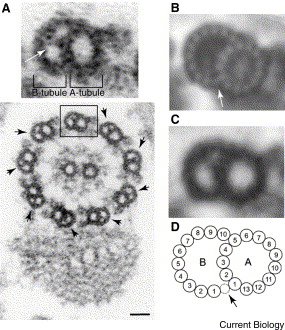
Centrosome are composed of two centrioles, barrel like structures made mostly of tubulin + other more unique proteins (see image below the fold). The number of centrosomes per cell is tightly regulated and the duplication of centrosomes is quite bizarre. You start off with a single centrosome. At some point (S phase?) the two centrioles come apart and new (or "daughter") centrosomes seem to grow off of the old (or "mother") centrosomes. Very reminiscent of semi-conservative replication of DNA. But it get stranger.

centrosome.jpg

If you watch the centrosomes throughout cell division, right before cytokinesis (the physical cleavage of two daughter cells) occurs, one of the two centrosomes from one of the two cells, migrates to the cleavage furrow and (in the words of Michel Bornens) gives it a "kiss". This event is thought to stimulate the final braking off of the two daughter cells.

Then about 5-6 years ago Alexey Khodjakov performed a remarkable experiment. He labeled the centrosomes with a green fluorescent protein (I think it was GFP-gamma-tubulin) then blasted them away with a laser. After centrosome duplication he blasted one of the two centrosomes and got cells with lopsided resulting mitotic spindles. The microtubules one side converged on a centrosome while the microtubules on the other side having no centrosome, became unorganized. Incredibly if he ablated both centrosome, the cells could form a spindle (mostly due to the fact that motor dynein can focus microtubules without centrosomes). The cells then progressed through mitosis and even divided (although sometimes they were trapped in cytokinesis - see Bornens' kiss). After division they got a huge surprize, the cells stoped dividing further. They discovered that centrosomes are needed to initiate a new round of DNA synthesis (or the G1-S transition). So just before DNA is copied, a cell asks itself "is my centrosome okay?" If the answer is yes => proceed with DNA copying. If not => stop cell division. You can advance through this "check point" if you get rid of p53, a key protein that regulates the cell cycle.

But why all the fuss? What is so special about the centrosome? Why are cells checking it? Why is it kissing stuff? Why?

Michel Bornens came up with this individuation theory. Centrosomes mark an individual cell. At certain points, cells check up on their centrosome to check on their individualistic identity. If something is wrong, such as improper cell division, cells can sense their non-individual state and activate programed death and/or other drastic measures. Screwed up cells (such as cancer cells) have all sorts of centrosome defects ... many have an overabundance of centrosomes and centrosome associated proteins suck as pericentrin.

And there's more! Many years ago, Lynn Margulis (famous for her theory of the endosymbiotic origin of mitochondria) proposed that centrosomes originated from the fusion of a spirochaete-like-organism (right, image of a spirochaete) and the prototypical eukaryotic cell. The spirochaete may have helped the host cell to move around (I never found this explanation very satisfying - the spirochaete must have offered some other benefit). In support of this idea, various publications in the 80s claimed that the centrosome contained its own nucleic acid, just like mitochondria and other products of symbiosis, but these experiments were never definitive.

Now out of Bob Palazzo's lab (no relation ... see extra comment at the end) Mark Alliegro's Lab (click here for a comment on this change) has come up with a truly remarkable finding. His group purified centrosomes from clam oocytes ... for some reason clams have huge centrosomes and thus makes this experiment a whole lot easier. Along with the centrosomes came ... certain specific RNAs. But wait, people have claimed this in the past, and one of the enriched RNA was 18s ribosomal RNA, perhaps it was contamination. On the other hand the enrichment was for only certain RNAs (five in this paper). Besides the 18s rRNA, the other four RNAs were not found in any genomic database. (For you RNA enthusiasts these have now been called cnRNAs - centrosomal RNAs.)

And one of these cnRNAs is QUITE intriguing. It encodes a protein that has similarity to 'RNA-dependent nucleotide polymerase'. Furthermore, this RNA probably gets translated into protein. (Warning: technical jargon comming up) The group maid antibodies against a peptide derived from the hypothetical protein, and found an immunoreactive band in lysates from oocytes and adult clam (end jargon).

So let me rephrase this. Clam centrosomes contain RNAs not found in any genome (so far), and one of these RNAs encodes an enzyme that could potentially copy RNA? Holly replication, Batman!

So could it be? Centrosomes have their own genome (an RNA genome at that?) Wow!

Ref:
Mark C. Alliegro, Mary Anne Alliegro, and Robert E. Palazzo
Centrosome-associated RNA in surf clam oocytes
PNAS (2006) 103:9034-9038


(Note: This whole Palazzo/centrosome thing is very quirky. He knows of me, I know of him, but we've never met. He has worked in the centrosome field for quite a while, I've published papers on a related structure, the microtubule organizing center. At meetings, some people think I have his long publication records, others who know Bob Palazzo well, ask me whether I'm his son -- he in fact does have a son named Alex. I almost went into the centrosome field by going to Steve Doxsey's lab (great guy!) but just couldn't live in Worcester. Now I'm studying (in part) RNA ... and Bob Palazzo is into cnRNA ... I know ... very strange.)

8.05.2006

RNA classification

Well two weeks ago in Science, two reports came out about yet another species of small RNA ... rasiRNA ... uhm i mean piRNA (OK they haven't harmonized their nomenclature yet).

So here is a brief review of the types of RNA:

- mRNA (messenger RNA). These are the RNAs that encode polypeptide chains.
- rRNA (ribosomal RNA). These form the core structure of the ribosome. The ribosome is the enzyme that translates the tri-nucleotides, to amino acids. In this way it synthesizes (or "translates") proteins from mRNAs.
- tRNAs (transfer RNA). These are used by the ribosome to translate the tri-nucleotie. On one end a loop displaying the complement of the tri-nucleotide is displayed, on the other end is the corresponding amino acids. So if you think about it tRNAs are responsible for coupling the tri-nucleotide to the amino acid.
- snRNA (small nuclear RNA). These form the core components of the splicesome, an enzyme that catalyses the splicing of mRNA.
- snoRNA (small nucleolar RNA). These strange critters help to process and assemble a variety of RNAs such as rRNAs.
- siRNA (small interfering RNA). These are processed from long double stranded RNAs, mostly exogenous in origin (exogenous = originates from outside the entity, in this case the cell) but occasionally are produced from endogenous transcripts (endogenous = originates from the same entity, in this case the cell's genome). The 21-23 nucleotide fragments are then used by the RNAi (RNA interference) pathway to turn off the expression of any single stranded RNA that the siRNA can base pair with. The machine that mediates RNAi is the RISC (RNAi Induced Silencing Complex), which includes the siRNA, a protein called Argonaute, and some other factors.
- miRNA (micro RNA). These are processed from short nearly double stranded small hairpin RNAs (shRNA) that are produced from endogenously transcribed RNAs. Like siRNAs, miRNAs are 21-23 nucleotides in length and are used by RNAi and the RISC machinery to inhibit mRNA transcription.
- rasiRNA (repeat associated small interfering RNA). These small RNAs (24-29 nucleotides in length) are produced from endogenous transcripts in fruit flies and seem to silence. Unlike siRNA, rasi RNA they are highly enriched in germline cells (sperm+oocyte) and form a complex similar to the RISC complex except the main protein involved is Piwi, a member of the Argonaute family of proteins. It seems like rasiRNAs function to inhibit bits of selfish DNA that copy themselves and multiply within the genome. These "selfish genes" are called retrotransposons.
- piRNA (piwi interacting RNA). Similar to rasiRNA, piRNAs are 25-31 nucleotides in length and produced from endogenous genes in mammalian cells. Along with Piwi, they form the piRNA complex (piRC) and act to. Unlike siRNA and miRNA, piRNA (and rasiRNA?) are not processed from a double stranded RNA precursor. For the moment the role of piRNAs is not clear, but like rasiRNA, these are highly enriched in germ cells. It is possible that they may regulate selfish genes, but there wasn't enough info in the paper ... I'm sure we'll find out soon what their targets are.

Ref:

rasiRNA
Vasily V. Vagin, Alla Sigova, Chengjian Li, Hervé Seitz, Vladimir Gvozdev, and Phillip D. Zamore
A Distinct Small RNA Pathway Silences Selfish Genetic Elements in the Germline
Science (2006) 313:320 - 324


piRNA
Nelson C. Lau, Anita G. Seto, Jinkuk Kim, Satomi Kuramochi-Miyagawa, Toru Nakano, David P. Bartel, and Robert E. Kingston
Characterization of the piRNA Complex from Rat Testes
Science (2006) 313:363 - 367

8.03.2006

Let's inhibit Cdc42

Secramine inhibits Cdc42-dependent functions in cells and Cdc42 activation in vitro.
HE Pelish, JR Peterson, SB Salvarezza, E Rodriguez-Boulan, JL Chen, M Stamnes, E Macia, Y Feng, MD Shair, and T Kirchhausen
Nat Chem Biol, Jan 2006; 2(1): 39-46

Cdc42 is a small GTPase associated with multiple cellular functions. It is "active" when is bound to GTP and membranes, and "inactive" when bound to GDP. This and other small GTPases are further regulated by RhoGDI, which regulates Cdc42 binding to membranes.

The paper I picked describes a small molecule - Secramine - that interferes with Cdc42-related activities, therefore it can be used as an inhibitor of Cdc42. The authors found that secramine does not interfere with the loading of GTP into Cdc42. Instead, secramine seems to interfere with membrane-dependent activation of Cdc42, probably by stabilizing the interaction between Cdc42 and RhoGDI. Further work is probably on the way to characterize the mechanism of action of secramine, but in the mean time the authors showed that secramine inhibited transport of NCAM from the golgi to the plasma membrane and golgi reorientation in migrating cells. These two cellular processes are known to be dependent on Cdc42. Secramine can be a very powerfull tool to use as a reversible inhibitor of Cdc42.