7.28.2006

Protein folding is coupled to mRNA stability ... across a membrane

Yes this is the surprising result interpretation of Jonathan Weissman's paper in Science. For non ER-aficionados, click here first, to get some background on the unfolded protein response (UPR) and ER associated degradation (ERAD). And to learn about some recent developments on ERAD, click here.

OK on to the HARDCORE cell biology ...

Remember under UPR conditions cells want to stop translating ER targeted proteins and instead synthesize chaperones and ERAD components. UPR inhibits translation through PERK (see the post on UPR) but what happens to the mRNA that encodes ER targeted proteins? Well it turns out that several of these mRNAs get degraded. This mRNA destruction requires IRE1 but not XBP1. If targeting of the mRNAs to the ER is disrupted, the mRNAs evade the UPR activated destruction.

So the question is why are certain ER targeted mRNAs destroyed (like those that encode plasma membrane or secreted proteins) while not others (like mRNAs that encode ERAD components)?

Well it could either be the mRNA that has a special code that makes it susceptible to UPR degradation, or the translational product may trigger the destruction of the mRNA.

To test the second idea, the Weissman group incorporate either a single, double or triple nucleotide insertion into the mRNA. Remember that a trinucleotide specifies an amino acid ... in the case of a single or double nucleotide insertion, the reading frame of every downstream trinucleotide is altered resulting in a completely different translational product. If a trinucleotide is inserted, the reading frame is conserved and all you've done is add an extra amino acid to your translational product. Well what they found was that when you altered the translational product, you abolished the mRNA's susceptibility to UPR mediated degradation. Yes the protein specifies the stability of the mRNA. The implication is that if a protein doesn't fold correctly as it is being translated and translocated into the ER, it targets the destruction of it's mRNA. But for the protein to direct the cleavage of the very mRNA it is coming from you would have to build a model where mRNA translation, translocation, and IRE1 dircted cleavage should all be happening at the same time (or in cis). Incredible! The model is nicely summarized in this figure from a David Ron review:

UPRmRNA2.gif
(click here for a larger version.)

One weird thing. By introducing a frame shift (i.e. insertion of 1-2 nucleotides in the coding region), the Weissman group transformed a natural protein into "junk" protein. This "junk" should have MORE problems folding than the original product, SO WHY IS IT NOT PROMOTING UPR MEDIATED mRNA DEGRADATION? (Very weird.)

This is a big finding, and there are some obvious experiments that could be done to give some more confidence to the model.

1) Alter an mRNA that isn't subjected to UPR mediated mRNA decay by adding a frame shift. The new mRNA will then encode "junk protein" that should have problems folding. By the new model, this new mRNA should be sensitive to UPR.
2) Test whether the mRNA destruction really operates in cis. This can be done by coexpressing two mRNAs, one with a frame shift and one without. If the expression of the frame shifted mRNA affects the stability of the unaltered mRNA then this process does not operate in cis (and hence in trans).

Overall a cool story.

Ref:
Julie Hollien and Jonathan S. Weissman
Decay of Endoplasmic Reticulum-Localized mRNAs During the Unfolded Protein Response
Science (2006) 313:104-107
7.25.2006

Actin differentiation through Arginylation

In cells, actin polymers dictate cell morphology.

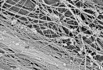
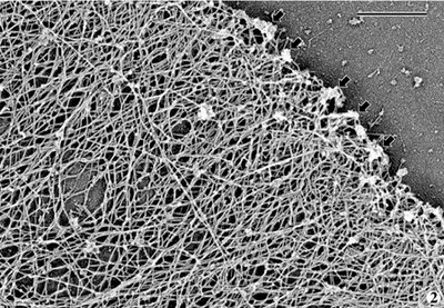
Actin filaments can adopt several conformations, they can be bundled into large microfilaments (often called stress fibers; here "mf" - electron micrograph taken from the Borisy lab webpage) or arranged in a meshwork (as seen in the second electron micrograph). One actin isomer (Gamma-actin) predominates in the stress fibers that are present in the cell body, while another (beta actin) predominates in the actin meshwork found in the leading edge of migrating cells. In fact, the generation of this meshwork right at the tip of the cell (see arrows in the first figure) is thought to push the cell membrane forward. This leading edge polymerization provides the major force for membrane spreading and plays an important role in cellular locomotion.

So if gamma-actin makes up microfilaments, and beta-actin makes up the meshwork, how do the two isoforms differ? How is the differential distribution explained? Beta-actin mRNA is concentrated at the leading edge and is thought to be synthesized there, but most researchers thought that there must be more to the story than that. Gamma-actin and beta-actin differ by only 4 out of 100 residues, and both genes are EXTREMELY well conserved across different species (100% conserved at the amino acid level between birds and humans).

A couple of weeks ago a great paper came out in Science about actin arginylation that cleared things up a bit.

For many years it had been noted that tubulin, the building block of the microtubule cytoskeleton, undergoes many post-translational modifications (see my post on this topic). These modifications help to differentiate microtubules that are aligned along the cell's main axis from the bulk microtubules that are present through out the cell. Now a group from U Penn who were studying arginine tRNA protein transferase (Ate1), discovered that actin gets post-translationally modified too. Bottom line - Ate1 adds arginine onto beta-actin's N-terminal glutamate side chain. This modification (similar to what was seen in tubulin & microtubules) aids in differentiating a part of the actin network from bulk actin filaments.

Now comes the new stuff ...

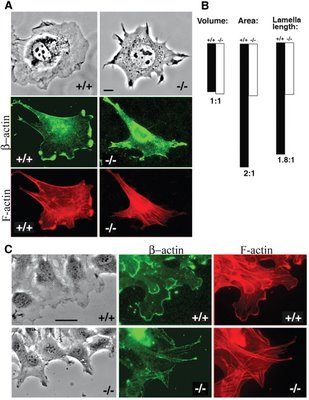
In the recent Science paper, the authors report that beta-actin, but not gamma-actin, gets arginylated by Ate1. This is supported by the finding that beta-actin arginylation is abolished in cells lacking Ate1. Aha! Perhaps arginine may give beta-actin the required properties to form meshworks. In agreement with this idea, Ate1 -/- cells have reduced levels of actin meshwork, and show defects in cell spreading and cell migration (see the figure, right). So then the big question is: how does arginylation affect beta-actin?


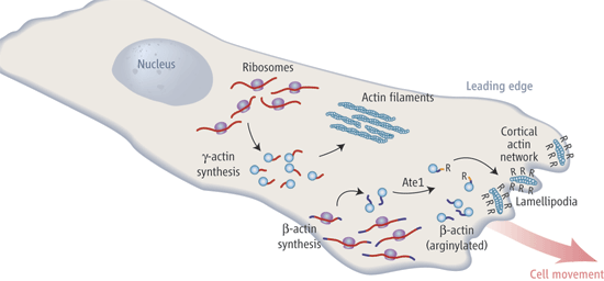
(here is a good schematic I ripped off of Chloe Bulinski's nice commentary in the same issue of Science).

The authors speculate that the extra positive charge from arginine may help prevent actin from bundling through electrostatic repulsion. Quite honestly I do not believe that the answer will be so simple. The actin meshwork and filaments are formed by separate mechanisms. The meshwork is formed by the polymerization of actin "branches" by the arp2/3 complex. Microfilaments are formed (probably) by formins which wrap around actin, forcing it into long fibers. These fibers are then bundled by the motor protein myosin. It is then likely that arginylation affects one of these actin-modeling processes.

In conclusion this paper offers a neat new concept for the cytoskeletal field.

Ref:
Marina Karakozova, Marina Kozak, Catherine C. L. Wong, Aaron O. Bailey, John R. Yates, III, Alexander Mogilner, Henry Zebroski, Anna Kashina
Arginylation of ß-Actin Regulates Actin Cytoskeleton and Cell Motility
Science (2006) 313:192-196

7.06.2006

Golgi Maturation

No soccer today. So instead of spending time watching others run around, go read the two papers, published in last week's Nature on Golgi maturation.

Proteins that need to traverse, or be embedded within membranes are synthesized in the endoplasmic reticulum (ER) and are transported cotranslationaly for the most part through a pore called the translocon. Most of these proteins are then delivered, through vesicular transport to the Golgi complex, where they are post-translationally modified. Sugars are added, sugars are removed ... indeed these proteins undergo many different modification steps. The Golgi is divided into many pancake shaped organelles stacked on top of each other, every pancake (or cisterna) having a different modification enzymes. Like penalty kicks and red cards, the Golgi seems to be an endless source of controversy. Does the Golgi dissolve into the endoplasmic reticulum during mitosis? Do proteins travel via vesicular transport from cisterna to cisterna, or does each cisterna "mature"?

ergolgi.jpg

But first, what does "mature" mean? It is the act of replacing one set of modifying enzymes with another set of modifying enzymes. So that early (or cis) Golgi is gradually transformed into Golgi containing the next set of modification enzymes (medial Golgi) which in turn is gradually transformed into late (or trans) Golgi. Well actually there are many cisterni (in the neighborhood of 6) in each Golgi stack. From the Golgi vesicles full of modified proteins are transported to various intracellular compartments such as the endosome recycling compartment.

How to tell whether Golgi cisterni "mature"? you would think that it's easy. Just fluorescently label proteins that traverse the Golgi and watch whether they stay in one cisterna or whether they travel across a stack. Unfortunately the stacks are smaller than the resolution limit of light microscopy. One could also image Golgi modification enzymes, but the size issue remains. Another attempted method, is to load the Golgi with giant particles. These substrates being too large to fit in vesicles, would presumably be stuck in one cisterna. But such experiments never gave conclusive evidence.

So what to do? Two groups solved this by reverting to yeast. (It seems like yeast are just simply the best organism to get at tricky problems ... ) You see yeast are weird. For a long time it was even questioned whether they had Golgi as no Golgi stacks were ever seen by electron microscopy. It turned out that the lack of stacks was due to the unique organization of the Golgi in yeast. You see in yeast the different cisterni are physicaly separated ... there are no stacks. And that is no problem for budding yeast, cuz they are so damn small. Also Golgi organization in "higher eukaryotes", is dependent on the microtubule network, but yeast do not use microtubules to direct vesicular transport. Now most proteins that are important for Golgi function are conserved between yeast and other eukaryotes, so even if ultra-structurally the Golgi shape may vary, the core operating system should be conserved.

OK fine so lets use yeast and image each separate Golgi sac to see if each sac matures, or to see whether substrates jump from sac to sac. To answer this, both groups labeled early and late Golgi markers and imaged the distribution of these markers over time. Do the markers jump from cisterna to cisterna? Or is each cisterna static in it's composition of modification enzymes.

So the answer: Golgi stacks mature. The rate of Golgi maturation is compatible with the rate of secreted protein production. If you block vesicle transport (via COP I temperature sensitive mutation) Golgi maturation slows down, indicating that modification enzymes get transported to and from each cisterna ... but since cistetna still matured, other transport mechanisms may help "mature" any particular Golgi sac.

OK that's all I have to say. Go check out the papers, and watch the movies. The results are pretty clear, hopefully the game on Sunday will provide for as much excitement.

Refs:

Eugene Losev, Catherine A. Reinke, Jennifer Jellen, Daniel E. Strongin, Brooke J. Bevis and Benjamin S. Glick
Golgi maturation visualized in living yeast
Nature (06) 441:1002


Kumi Matsuura-Tokita, Masaki Takeuchi, Akira Ichihara, Kenta Mikuriya and Akihiko Nakano
Live imaging of yeast Golgi cisternal maturation
Nature (06) 441:1007


Crossposted at The Daily Transcript.