6.29.2006

Stochastic expression of proteins in a single cell

Every thought about how variable the expression of a particular gene is across an entire cell population?

That’s what the Weissman lab described in a manuscript in a recent issue of Nature. Anytime you want to take on such a project – take my advice, you turn to yeast. The yeast field has created a library of strains, each containing a copy of GFP (Green fluorescent protein) fused to a particular gene within the genome. If you measure the fluorescence, you can quantify the level of protein expression. The next trick is to use flow cytometry to rapidly measure the brightness individual cells in a population. Brightness per cell = GFP per cell = expression of the tagged gene, per cell.

Now you can record how a particular gene is expressed, in terms of protein levels, on a cell to cell level. In addition one can identify how cells alter their protein levels when exposed to various conditions. Note that this type of experiment has been done at the mRNA level using microarrays, yet until now no one has published any account of how to perform these measurements at the protein level.

So what did they find?


- For cells grown in rich media, 30% of their genes have elevated protein expression, 10% have decreased protein expression when compared to cells grown in minimal medium. Proteins with increaed levels in rich media, are those involved in cell division and cell wall biosynthesis. Proteins with increaed levels in minimal media, are biosynthetic genes (if the environment doesn’t have aminoacids and nucleotides, you’ve got to make them yourself.)
- Importantly, the researchers asked whether protein levels reflected mRNA levels, or whether other post-translational events (such as protein degradation) played significant roles. To the relief of many microarray manufactures, changes in protein levels largely correlated with changes in mRNA fir most genes. There were some exceptions (like ribosomal RNA processing enzymes and enzymes involved in the Kreb cycle).

But now comes the interesting part, proteins involved in core functions (i.e. ribosomal proteins, initiators of translcription, protein synthesis, protein degradation) have low variability in protein levels on a cell-to-cell basis. The output from these genes had low noise. Strangely, Golgi components have low noise as well, but mitochondrial and peroxisome proteins have high noise. Overall it seems like mRNA production is the greatest determinant of noise:

…variation most likely originates from the stochastic production and destruction of mRNA molecules. Indeed, the magnitude of the variation observed here (CV 30% for low–medium abundant proteins) is entirely consistent with that expected if protein variation results from Poisson noise owing to small mRNA numbers (1–2 per cell) and is mitigated by a filtering effect that arises because proteins are typically far longer-lived than their messages.

...

... high noise is likely to be due, at least in part, to the introduction of a slow step into the production of mRNA, making the process more prone to bursts.


Furthermore, noisy genes are regulated at the transcriptional level by similar transcription factors and chromatin remodeling enzymes. Stable genes tend to be regulated by another group of transcription factors. Stable genes are also less likely to be affected by fluctuating mRNA numbers. This is best achieved by have increased numbers of messages that turnover rapidly.

The authors point out that noise (or the variability of expression) is an important consideration in how genes are regulated. Cells may want certain genes, such a those that respond to environmental stress, to have “noisy outputs”.
for some proteins that permit cells to respond to environmental perturbations, excursions from the mean at the single-cell level might benefit populations. In the short term, such deviations might facilitate a cell's initial response to environmental variation. More generally, the capacity to vary might permit a population to sample multiple phenotypic states to maximize the chances of some, but not all, cells' survival in an adverse environment.

Other genes, such as those involved in ribosomal maintenance and cell cycle regulation, need to have stable levels in order to ensure cellular homeostasis.

The takehome message, forget about the average, the generation of variability or stability may be a crucial component to how a cell is hardwired.

Ref:
John R. S. Newman, Sina Ghaemmaghami, Jan Ihmels, David K. Breslow, Matthew Noble, Joseph L. DeRisi and Jonathan S. Weissman
Single-cell proteomic analysis of S. cerevisiae reveals the architecture of biological noise
Nature (2006) 441:840-846


Cross posted at The Daily Transcript.
6.26.2006

Giving your mRNA a boost by increasing GC content

Abstract from PLoS (the abstract says it all):

Mammalian genes are highly heterogeneous with respect to their nucleotide composition, but the functional consequences of this heterogeneity are not clear. In the previous studies, weak positive or negative correlations have been found between the silent-site guanine and cytosine (GC) content and expression of mammalian genes. However, previous studies disregarded differences in the genomic context of genes, which could potentially obscure any correlation between GC content and expression. In the present work, we directly compared the expression of GC-rich and GC-poor genes placed in the context of identical promoters and UTR sequences. We performed transient and stable transfections of mammalian cells with GC-rich and GC-poor versions of Hsp70, green fluorescent protein, and IL2 genes. The GC-rich genes were expressed several-fold to over a 100-fold more efficiently than their GC-poor counterparts. This effect was not due to different translation rates of GC-rich and GC-poor mRNA. On the contrary, the efficient expression of GC-rich genes resulted from their increased steady-state mRNA levels. mRNA degradation rates were not correlated with GC content, suggesting that efficient transcription or mRNA processing is responsible for the high expression of GC-rich genes. We conclude that silent-site GC content correlates with gene expression efficiency in mammalian cells.


Very Interesting. High GC content at codon position #3, led to higher protein content, higher mRNA content, but no change in mRNA degradation. Unfortunately the authors did not test whether the mRNAs were transcribed more efficiently. Other possibilities is that mRNA 3' processing is inefficient or mRNA export is compromised (with the unexported mRNA being degraded immediately).

It's something to think about when designing your own genes. This finding also has implications for biologists who track mutations at synonymous sites ... although these mutations may be "silent" in how they change the coding of a protein, they may not be so silent (different use of the term) with regards to how they affect protein expression. This may also be a way of generating variability with regards to protein expression between different alleles. On the other hand, the effects of one point mutation may be very small. Is it too small to be selected on? We'll see.

Kudla G, Lipinski L, Caffin F, Helwak A, Zylicz M (2006) High Guanine and Cytosine Content Increases mRNA Levels in Mammalian Cells. PLoS Biol 4(6): e180

Cross posted at Daily Transcript.

6.21.2006

RNA decay Particles

Ujwal Sheth from Roy Parker's lab details the molecular mechanism that targets RNAs with premature stop codons to processing-bodies (or p-bodies) via the non-sense mediated decay (NMD) pathway. P-bodies are dense cytoplasmic granule-like structures that serve as sites of mRNA storage/degradation. P-bodies contain decapping enzymes, RNAses and many other proteins of unkown function. In this paper the authors demonstrate that the NMD component, and RNA helicase, Upf1p, targets aberrant mRNA to granules. Upf1p's ATPase activity is then required to recruit Upf2p and Upf3p to p-bodies.

Ujwal Sheth and Roy Parker, Targeting of Aberrant mRNAs to Cytoplasmic Processing Bodies.
Cell (2006) 125:1095-1109


It remains unclear how premature stop codons are recognized in yeast. In higher eukaryotes, if a stop codon occurs before a splice site, ribosomes fall off the RNA before they can kick off exon junction complexes that mark sites along the RNA where splicing has occurred. The exon junction complex then recruits NMD components that target the mRNA for destruction.

There has been much fuss lately with these p-bodies and the related structures termed "stress granules". Both structures are seen in most eukaryotes and play several seemingly incompatible roles. In general non-translating cytosolic mRNAs are shuffled into these structures. But why?

Some facts about RNA bodies:
- these cytosolic structures do not contain membranes yet are very dense and exclude large proteins
- much of the maternal RNA in oocytes is stored in granules
- a related structure, termed simply "RNA granules", transport RNA up dendrites in neurons
- stress granules are thought to be formed by the aggregation of TIA1, a protein thought to have prion activities
- neuronal RNA granules are thought to be regulated by CPEB (Cytioplasmic Polyadenylation Element Binder), another protein thought to have prion properties
- RNAi components target RNAs to p-bodies, and proteins involved in RNAi are enriched in p-bodies

The question is, why pack RNA so tightly into dense structures? And why form these particles with aggregating prions? Prions can exist in several forms, so perhaps RNA granules must adapt several roles? Some RNA granules, such as those in neurons and oocytes store RNA (i.e. a precious cargo), but in other cases RNA granules act as trash compactors. I'm sure that this story will get more interesting in the coming months/years.

6.09.2006

ScienceSampler


Myosin VI is an unusual motor

Myosin VI Stabilizes an Actin Network during Drosophila Spermatid Individualization

Tatsuhiko Noguchi, Marta Lenartowska, and Kathryn G. Miller
Mol. Biol. Cell 2006 17: 2559-2571


Similar to other myosins, myosin VI contains an ATP-dependent motor domain, a coiled-coil domain, and a globular tail domain. However, myosin VI moves towards the pointed end as opposed to the barbed end of an actin filament. Previous work has implicated myosin VI as a motor for endosomal movement due localization studies and in vitro assays showing the ability for myosin VI to form dimers and move processively. However, myosin VI can also act as an actin dependent molecular crosslinker. When expressed in baculovirus, a majority of myosin VI is monomeric and shows no processive movement.

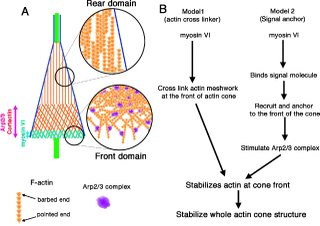
This paper sheds light on the role of myosin VI in vivo during spermatogenesis in drosophila, specifically in the actin cone. The deletion of myosin VI decreases both the relative amount and density of F-actin as opposed to wild-type in actin cones, while overexpression of myosin VI has the opposite effect. These data combined with the effect of myosin VI deletion on actin cones by EM, and the persistence of GFP-myosin VI after FRAP, illustrates myosin VI's role as a crosslinker. The above cartoon shows, a role for myosin VI stabilization of the branched actin meshwork at the front of actin cones. No data obtained from this paper implicates myosin VI as a cargo transporter.

Additionally, the structure of the actin cone is unique as the filaments in the cone are oriented with their barbed ends facing away from the direction of movement. This mechanism is opposite of that found in Listeria comet tails and lamellipodia where barbed ends face toward the direction of movement. This difference causes the authors to speculate on the actin polymerization mechanism which is also depicted in the cartoon.

6.07.2006

Why do baby neurons need LIS1?

This time I picked an "old" paper. It was published last year on JCB and describes the effects of knocking down Lis1 during brain development.

LIS1 RNA interference blocks neural stem cell division, morphogenesis, and motility at multiple stages.
Tsai JW, Chen Y, Kriegstein AR, Vallee RB.
J Cell Biol. 2005 Sep 12;170(6):935-45


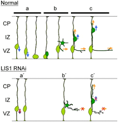
Cortical neuron migrate from the ventricular region to the perifery of the brain during brain development. Cortical neurons originate from the division of radial glial cells. Prior to cell division, the nucleus from radial cells migrates, away from the ventricular region, then returns to the initial position. Only after this detour, known as nuclear oscilation, radial glial cells divide. The daugther cells migrate to the sub-ventrical zone where they stop and become multipolar. Then they start extending axonal processes, become bipolar, and resume migration.
Lis1 mutations originate Lyssencephaly. Pacients with this mutations have severe defects on brain development and die 1-2 years after birth. Lis1 mutations are thought to interfere with neuronal migration required for brain development.
In this paper, the authors provide clear evidence for a role of Lis1 in different stages of neuronal migration. They used a very powerfull technique (in utero electroporation) to introduce fluorescent siRNA or shRNA targeting Lis1 into cells at the ventricular region. Then they analised the position and shape of transfected cells, and most impressivelly, they followed neuronal migration in slices by timelapse microscopy, up to 18h.
Using these approached they show that Lis1 is required for nuclear oscilations and cell division in astral glial cells, bipolar migration and axonal extension.